

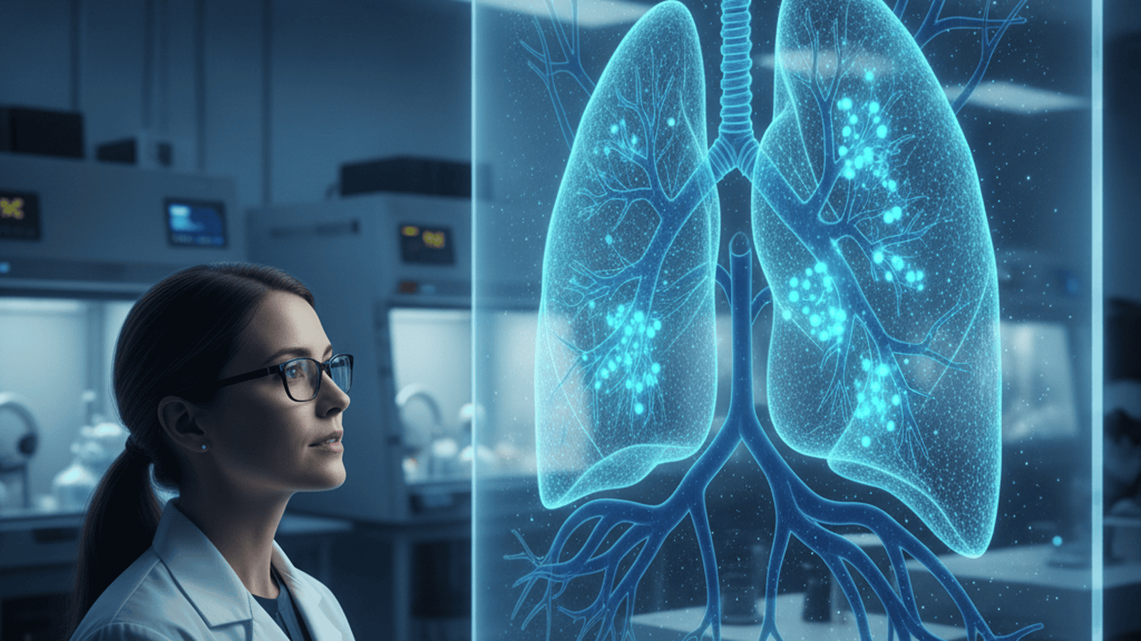
Researchers at the University of Pennsylvania have identified a unique population of “pioneer” cells that appear in the lungs following severe injury, such as viral pneumonia. These cells are essential for rebuilding the delicate air-sacs required for oxygen exchange, offering a potential new target for regenerative medicine.
TLDR: Scientists have discovered a specific state of lung cells that acts as a “pioneer” to repair damage from severe respiratory infections. By identifying the signals that trigger these cells, researchers hope to develop therapies that accelerate recovery for patients with chronic lung disease or post-viral complications like pulmonary fibrosis.
Researchers at the University of Pennsylvania’s Perelman School of Medicine have identified a specialized population of cells that play a critical role in repairing the lungs after severe injury. These “pioneer” cells, a specific state of alveolar epithelial cells, appear to be the primary drivers of regeneration in the delicate tissues where oxygen enters the bloodstream. The discovery provides a new understanding of how the body recovers from conditions like severe influenza and COVID-19, offering a potential roadmap for future regenerative therapies. This breakthrough is particularly timely as the global medical community continues to grapple with the long-term respiratory consequences of the recent pandemic.
The study focused on the alveoli, the tiny air sacs that facilitate gas exchange. When these structures are damaged by viral pneumonia or acute respiratory distress syndrome (ARDS), the body must rapidly rebuild the epithelial lining to prevent permanent scarring or respiratory failure. The research team, led by experts in regenerative medicine, utilized advanced single-cell RNA sequencing to track the lineage and behavior of cells during the recovery phase in both animal models and human tissue samples. This high-resolution mapping allowed the scientists to observe cellular transitions in real-time, revealing a complexity previously hidden by traditional microscopy techniques.
The findings revealed that a subset of cells, which the researchers termed “pioneer” cells, emerge from existing Alveolar Type 2 (AT2) progenitor populations. These cells exhibit a unique genetic signature, characterized by the expression of Keratin 8 (KRT8), which allows them to migrate into damaged areas more effectively than standard lung cells. Once they reach the site of injury, they act as a biological scaffold, signaling other cells to follow and eventually differentiating into the mature Type 1 cells necessary for gas exchange. This “pioneer” state is temporary but essential; without it, the structural integrity of the lung cannot be restored, leaving the organ vulnerable to further collapse.
A key component of this process is the activation of specific signaling pathways, including the Wnt and Notch pathways, which dictate the timing and scale of the repair. The researchers observed that in cases of chronic lung disease or severe aging, these pioneer cells often become “stuck” in an intermediate state. Instead of completing the repair process and transitioning into functional lung tissue, they remain in a pro-inflammatory state that contributes to the formation of fibrotic tissue. This fibrosis stiffens the lungs and significantly reduces their efficiency, a hallmark of many chronic respiratory conditions. The study suggests that the environment of the damaged lung—the “niche”—plays a decisive role in whether a pioneer cell successfully rebuilds the tissue or contributes to a permanent scar.
This distinction between successful regeneration and pathological scarring is a major focus for the medical community. By identifying the molecular triggers that keep pioneer cells on the path toward healthy repair, scientists may be able to develop drugs that prevent the onset of pulmonary fibrosis. Such treatments would be particularly beneficial for patients who suffer from long-term respiratory impairment following intensive care hospitalization or those with idiopathic pulmonary fibrosis. The ability to pharmacologically nudge these cells toward a regenerative fate could revolutionize post-viral care and improve the quality of life for millions of survivors.
The research also highlights the remarkable plasticity of the human respiratory system. Previously, it was thought that lung repair was a more static process involving only a few types of resident stem cells. The identification of a dynamic, transitional “pioneer” state suggests that the lung has a more sophisticated and flexible toolkit for responding to environmental threats than previously understood. This flexibility allows the organ to prioritize immediate structural integrity before returning to its specialized gas-exchange functions. It represents a shift in how biologists view organ recovery, moving from a model of simple replacement to one of complex, multi-stage reconstruction.
Future studies will aim to determine if these pioneer cells can be stimulated exogenously through targeted molecular therapy. If clinicians can jumpstart the production of these cells in patients with failing lungs, it could reduce the duration of mechanical ventilation and improve survival rates for ARDS. The team is currently screening chemical compounds that might enhance the natural regenerative capacity of these newly discovered cellular architects, moving closer to a reality where lung damage is no longer a permanent sentence. This work underscores the importance of basic cell biology in solving some of the most pressing challenges in modern clinical medicine.

Susan Carter serves as a Senior Correspondent for Just Right News, where she leads the network’s comprehensive coverage of Health, Medicine, and Public Policy. With a career dedicated to dissecting the complexities of the American healthcare system, Susan brings a principled perspective to the most pressing debates of the day. Her reporting is characterized by a commitment to individual liberty, fiscal responsibility, and a healthy skepticism of government overreach, making her a vital voice for audiences seeking clarity in an increasingly regulated landscape.
Raised in Minneapolis, Minnesota, Susan’s professional outlook is deeply informed by the values of the American heartland. Growing up in the Twin Cities, she witnessed firsthand the importance of community-driven solutions and the resilience of the individual. This Midwestern foundation instilled in her a firm belief that the best answers to public policy challenges often come from local innovation and personal responsibility rather than top-down federal mandates. Her background allows her to translate complex legislative jargon into stories that resonate with families who value common sense and local autonomy.
Now based in Baltimore, Maryland, Susan operates from a unique vantage point at the intersection of medical innovation and urban policy. Living and working in a city renowned for its world-class medical institutions and significant public health challenges, she has a front-row seat to the realities of the modern healthcare landscape. Her proximity to these hubs of research and policy allows her to scrutinize how government intervention affects the quality of care and the freedom of medical professionals. In Baltimore, she sees the tangible results of policy decisions, using the city as a lens through which to examine the broader national health debate and the impact of federal spending on local communities.
As the lead for the acclaimed feature series “The Cost of Care,” Susan dives deep into the economic realities facing patients and providers alike. Her work focuses on the necessity of price transparency, the benefits of market competition, and the preservation of the sacred doctor-patient relationship. She is a staunch advocate for the idea that a well-informed public is the best defense against bureaucratic inefficiency. Through this series, she has exposed the hidden drivers of rising medical expenses, always seeking to empower the individual consumer over the massive institution.
Susan’s reporting on public policy is characterized by a commitment to rigorous inquiry and a focus on constitutional limits. Whether she is analyzing new pharmaceutical regulations, investigating the impact of federal health mandates, or exploring the future of medical technology, her goal remains the same: to provide Just Right News audiences with the facts they need to navigate a complex world. By blending her Midwestern sensibilities with the insights gained from her base in Maryland, Susan Carter has become a trusted authority for those who believe that personal freedom and public health are inextricably linked. Her work continues to challenge the status quo, advocating for a system that respects the taxpayer and prioritizes the patient.